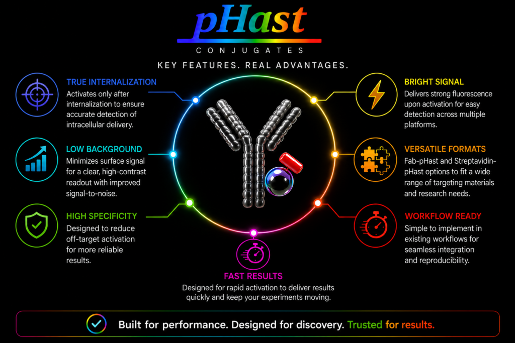
Internalization data is only useful if you can trust what you’re seeing.
Because pHast Conjugates activate specifically in acidic intracellular compartments, they help distinguish internalized material from surface-bound signal and reduce the background that can complicate interpretation with traditional assays.
The result is a clearer, more biologically relevant readout of internalization with less ambiguity and less assay complexity.
Saporin as the Scalpel in Molecular Surgery to create Models of Dementia, Alzheimer’s Disease and Parkinson’s Disease

How to explore
Advanced Targeting Systems
Products
Filter Products By:

Library


Featured


ZAP Kits
Which kit is right for your research? Check out this video or browse our list of ZAP products.
Secondary Antibodies
Browse our selection of Secondary Antibodies providing reliable, consistent performance across a wide range of research applications


Have a question about your next project?
Contact us and let’s get started targeting together.